Coral Taphonomy
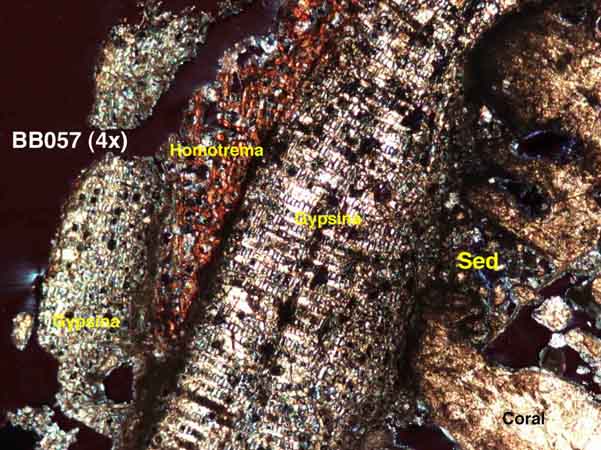
The above photos is a photomicrograph (2X) of a crust
on a branch of standing Acropora palmata that was killed by White
Band Disease in the late 1970s. The colony was still standing in January
2002. Note the importance of Gypsina sp. and Homotrema rubrum.
Also inportant on other samples are coralline algal crusts and vermetid
gastropods. Crusts on upper branch surfaces appear to be different than
those on the lower side of the same sample.
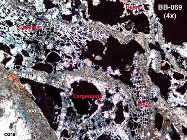
The above photo is a photomicrograph (2X) of an A.
palmata branch collected from within an excavation into the reef
substrate at the same site. Note how much "lacier" the crusts
look, owing to a much greater abundance of the foram Carpenteria
spp. The more open framework of the encrusters and the more complex sequences
is thought to reflect less grazing among the broken branches piled up
by storms.
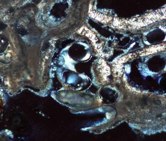
Photomicrigraph (10X) of worm tubes in a crust from
one of the excavated samples from the reef interior.
Where are we?
-
All samples have been slabbed, impregnated and thin sectioned.
- Gestalt differences in crusts associated with storm deposition versus standing, dead colonies have been identified.
- We are starting the process of gethering quantitative data from the samples. This is largely being accomplished by Oberlin undergraduate students.
- Anyone interested in collaboration can contact me at "dennis.hubbard@oberlin.edu". These are few of us looking at encrusting organisms and we can use all the help we can get.